
MEASUREMENT
Quantify geometry
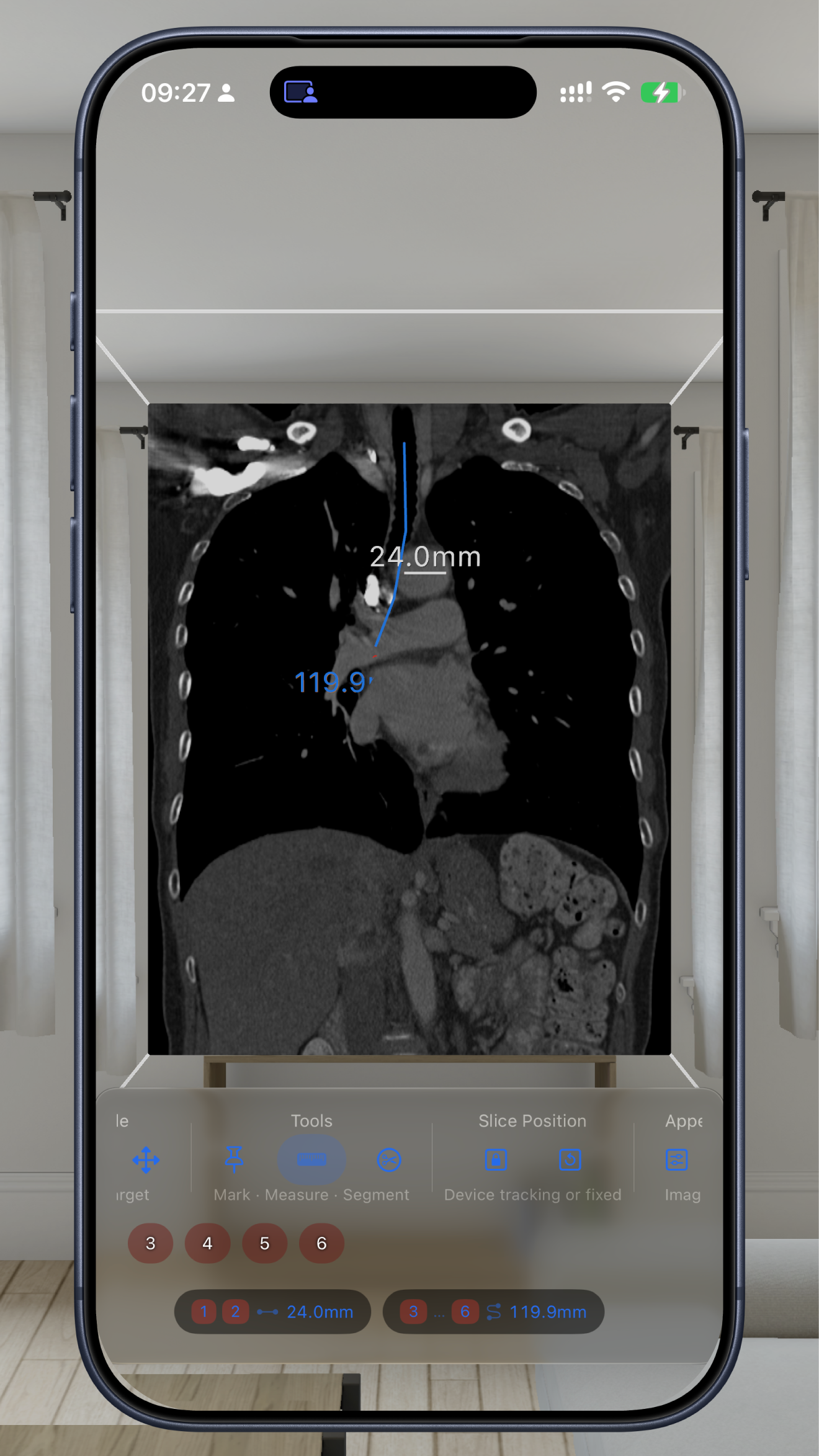
Measure distances, angles, and paths directly in 3D. Export measurements as CSV for further analysis.
Volumetric medical imaging has always been three-dimensional. Now the interaction is too.
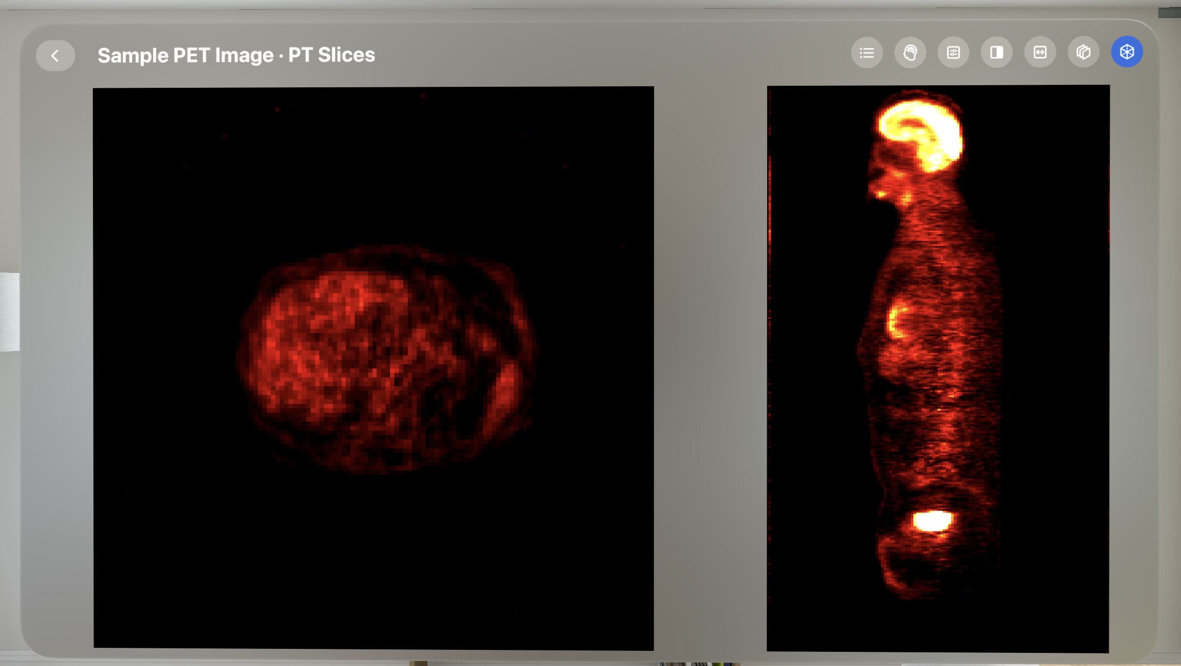
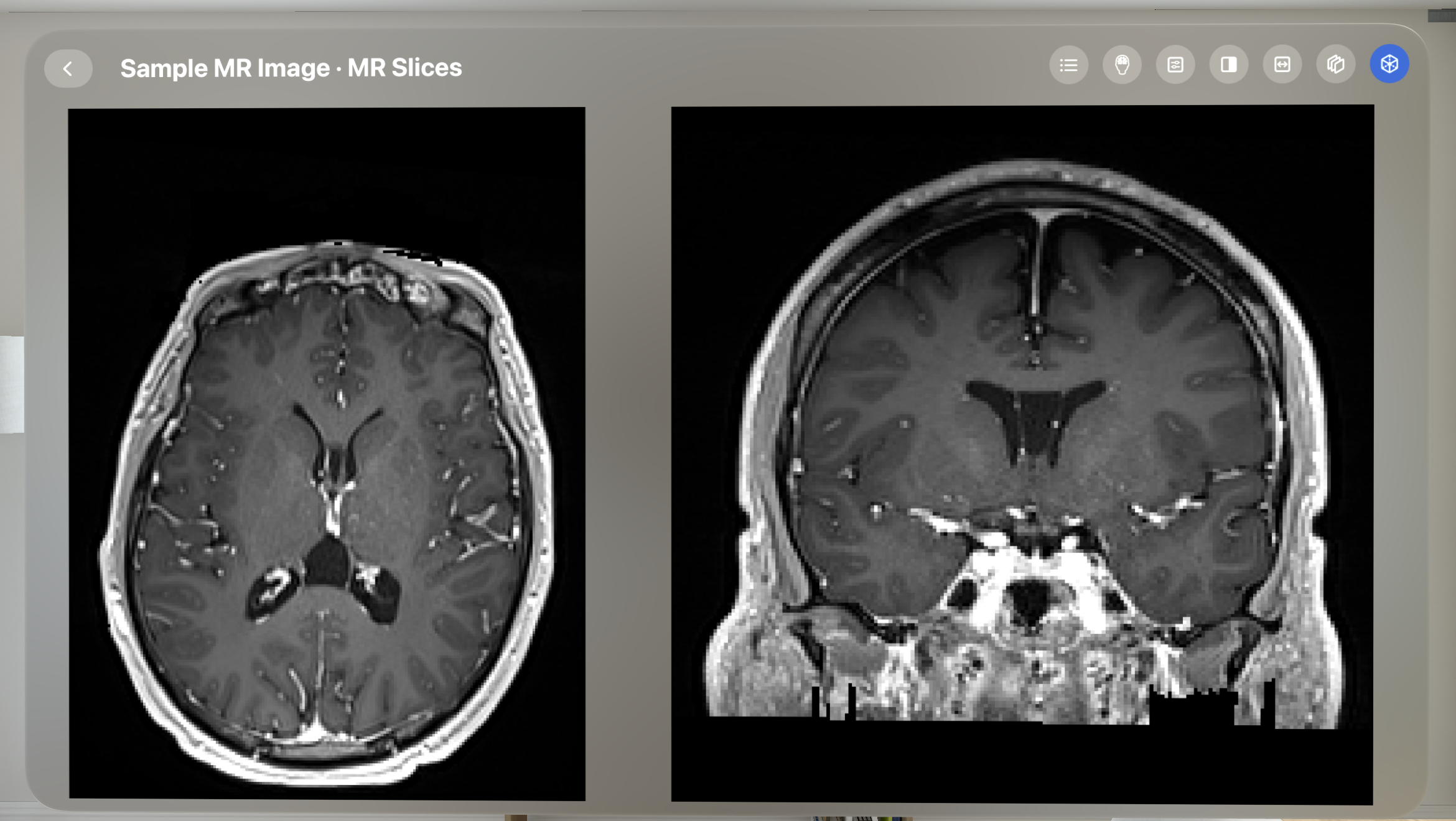
Measure, segment, and register in 3D on Apple Vision Pro, iPad, and iPhone.
Measure distances, angles, and paths directly in 3D. Export measurements as CSV for further analysis.
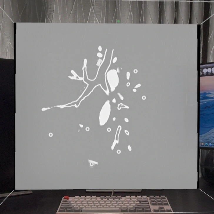
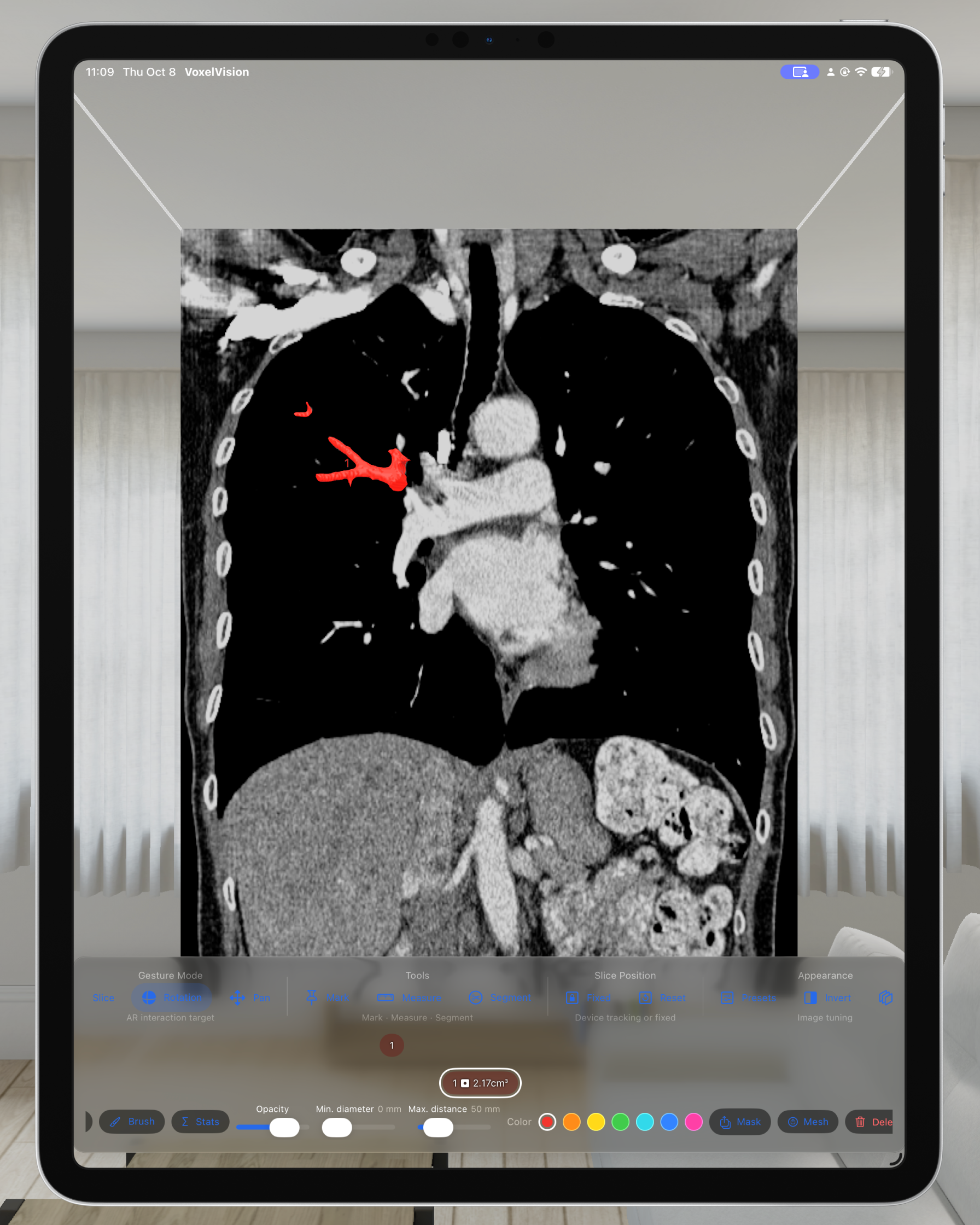
Separate regions of interest and refine their boundaries. Export labeled volumes, 3D surfaces, and statistics for further study.
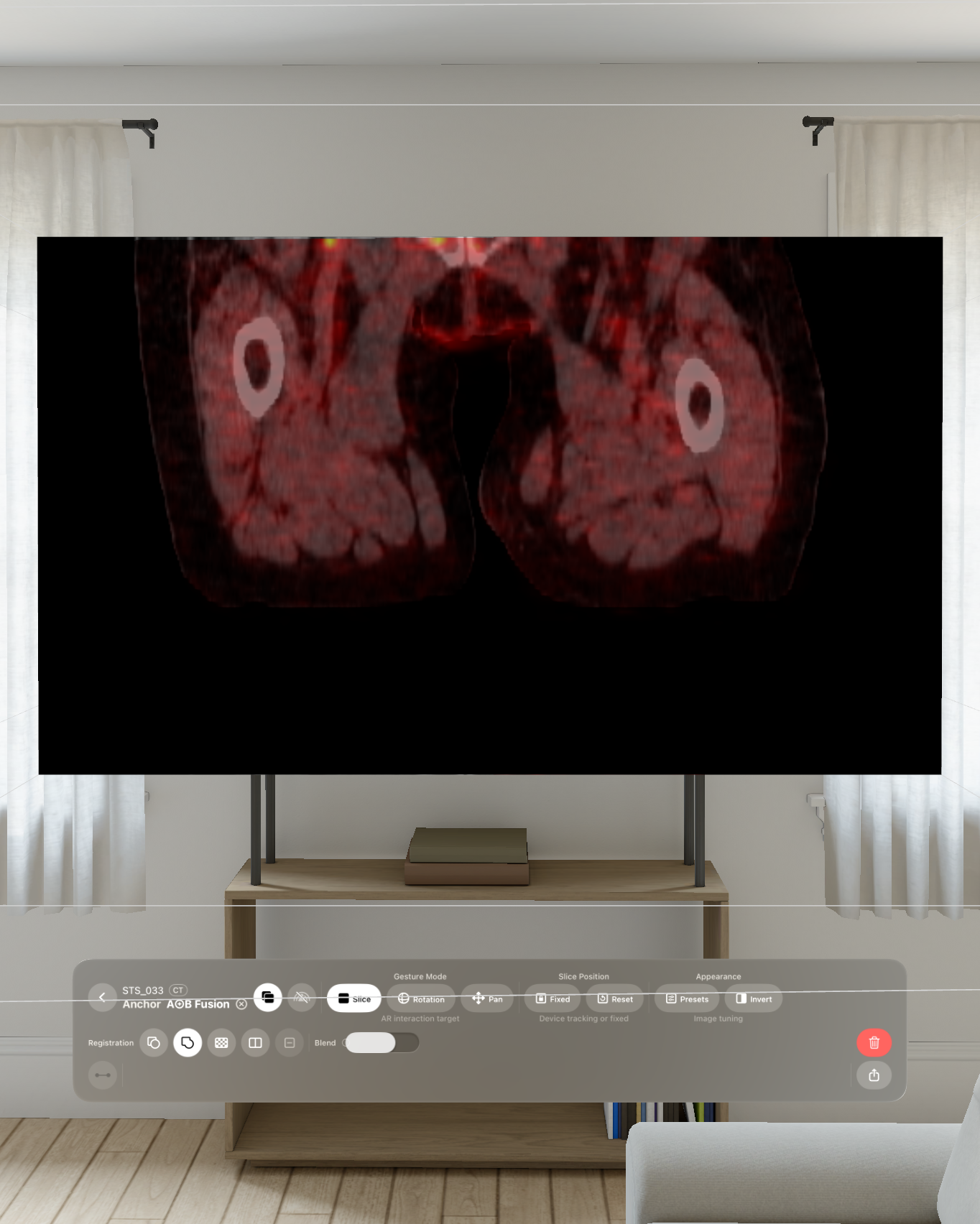
Bring two scans into alignment and compare them in a fused view. Save the alignment for your workflow.
The same tools are available on all three platforms. Tools are free to try on sample images; in-app purchases unlock them for your own cases.
Explore the same case together with SharePlay. Invite others to view or edit, and choose whether to keep the session's annotations.
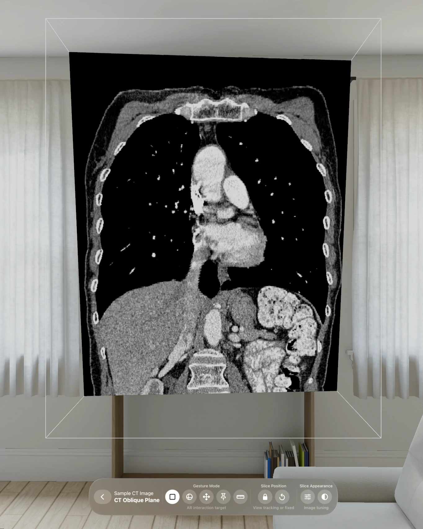
Follow curved structures, view thicker slices, and inspect image intensity. Adjust image contrast and PET colors to explore the details.
Work through planned scan pairs and process saved alignments in batches. Export results and step-by-step records for repeatable research workflows.
Use the same spatial interaction as you measure, segment, and register. Follow your viewpoint, or position the plane by hand.
The oblique plane follows your viewpoint — head-tracked on Apple Vision Pro, device-tracked on iPad and iPhone. Move yourself, and the cut moves through the anatomy.
Lock the plane in place and drag it through the volume by hand — for slow, deliberate positioning and repeatable views.
Try both interaction modes for free with the sample images.
In addition to standard DICOM, VoxelVision supports NIfTI and NRRD formats.
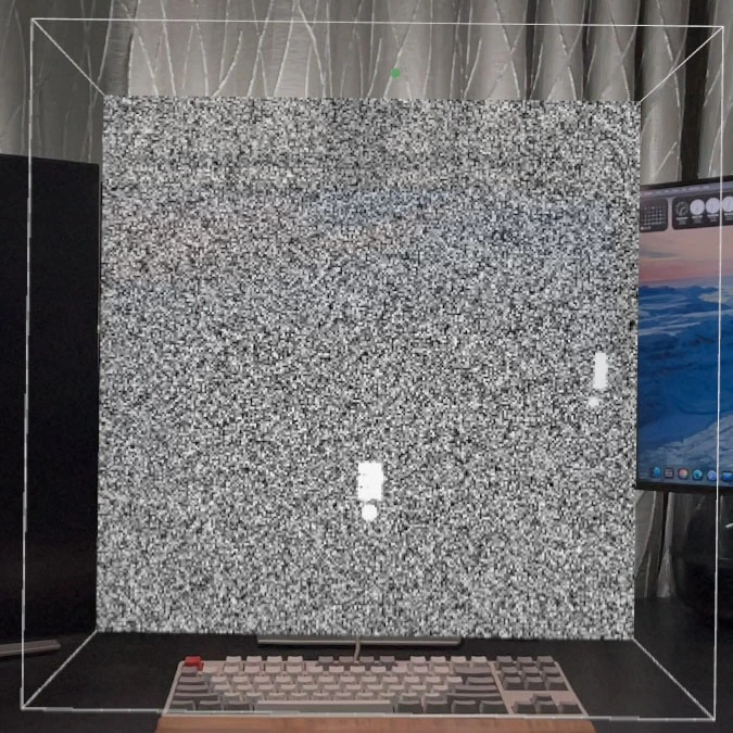
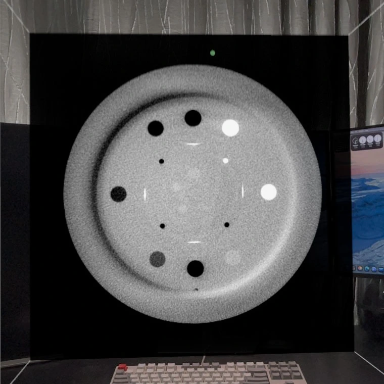
How do spatial measurements compare with known dimensions? A study using VoxelVision 1.2.1 tested distances, angles, paths, and perimeters on reference objects, with 570 blinded measurements across 100 volumes.
This study evaluated measurement with a single trained operator on reference objects, not segmentation, registration, or performance on clinical anatomy. VoxelVision is for education, research, and workflow exploration, not clinical diagnosis or treatment.
The study characterized pose-tracked double-oblique multiplanar reformation across three types of reference phantom, on head-mounted and handheld platforms.
Across 570 blinded measurements on 100 single-target volumes, the synthetic-phantom results were statistically equivalent across voxel spacing, platform, and session re-entry. Characterized with a single trained operator on phantoms with known reference geometry; generalization across independent operators and extrapolation to clinical anatomy define the boundary for subsequent study. VoxelVision is not a certified medical device.
Manuscript currently in peer review · full citation with doi to be added upon publication.
Open code & data — analysis code, measurement records, and per-tier execution lists: github.com/mingweicui/VV-measurement-validation (MIT). Author-built phantom datasets (MCVV-SPH, MCVV-RPH) archived on Zenodo (CC-BY-4.0).